How is mesothelioma diagnosed?
A diagnosis of mesothelioma is most often obtained with careful assessment of clinical and radiological findings in addition to a confirming tissue biopsy. (Learn about typical mesothelioma symptoms.) A review of the patient's medical history, including history of asbestos exposure is taken, followed by a complete physical examination, x-rays of the chest or abdomen, and lung function tests. A CT scan or MRI may also be done at this time. If any of these preliminary tests prove suspicious for mesothelioma; a biopsy is necessary to confirm this diagnosis.
Imaging Techniques and Their Value in Diagnosing and Assessing Mesothelioma
There are several imaging techniques which may prove useful when mesothelioma is suspected due to the presence of pleural effusion combined with a history of occupational or secondary asbestos exposure. While these imaging techniques can be valuable in assessing the possibility of the cancer, definitive diagnosis is still most often established through fluid diagnosis or tissue biopsy.
Some of the most commonly used imaging methods include:
• X-ray
A chest x-ray can reveal pleural effusion (fluid build-up) which is confined to either the right (60%) or left (40%) lung. On occasion, a mass may be seen. Signs of prior non-cancerous asbestos disease, such as pleural plaques or pleural calcification, or scarring due to asbestosis may also be noted.
• Computed Tomography (CT)
CT scans are also able to define pleural effusion, as well as pleural thickening, pleural calcification, thickening of interlobular fissures, or possible chest wall invasion. CT, however, is not able to differentiate between changes associated with benign asbestos disease (pleural disease), or differentiate between adenocarcinoma of the lung wh
ich may have spread to the pleura verses mesothelioma. CT scans may also be valuable in guiding fine needle aspiration of pleural masses for tissue diagnosis.
• Magnetic Resonance Imaging (MRI)
MRI scans are most often used to determine the extent of tumor prior to aggressive treatment. Because they provide images in multiple planes, they are better able to identify tumors as opposed to normal structures. They are also more accurate than CT scans in assessing enlargement of the mediastinal lymph nodes (those lymph nodes which lie between the two lungs), as well as a clear diaphragmatic surface, both of which play an important role in surgical candidacy.
• Positron Emission Tomography (PET)
PET imaging is now becoming an important part of the diagnosis and evaluation of mesothelioma. While PET scans are more expensive than other types of imaging, and are not always covered under insurance, they are now considered to be the most diagnostic of tumor sites, as well as the most superior in determining the staging of mesothelioma. Further explanation of PET scans.
• CT/PET
For patients who may be candidates for aggressive multimodality treatment (surgery, chemotherapy and radiation), accurate clinical staging is extremely important. Integrated CT/PET imaging provides a relatively new tool in this respect, and has become the imaging technique of choice for determining surgical eligibility. By combining the benefits of CT and PET (anatomic and metabolic information) into a single scan, this technology can more accurately determine the stage of the cancer, and can help identify the best treatment option for the patient. Read about a study of CT-PET imaging in preoperative evaluation of patients with malignant pleural mesothelioma.
A needle biopsy of the mass, or the removal and examination of the fluid surrounding the lung, may be used for diagnosis, however, because these samples are sometimes inadequate as far as determining cell type (epithelial, sarcomatous, or mixed) or because of the unreliability of fluid diagnosis, open pleural biopsy may be recommended. In a pleural biopsy procedure, a surgeon will make a small incision through the chest wall and insert a thin, lighted tube called a thoracoscope into the chest between two ribs. He will then remove a sample of tissue to be reviewed under a microscope by a pathologist. In a peritoneal biopsy, the doctor makes a small incision in the abdomen and inserts a peritoneoscope into the abdominal cavity.
Once mesothelioma is suspected through imaging tests, it is confirmed by pathological examination. Tissue is removed, put under the microscope, and a pathologist makes a definitive diagnosis, and issues a pathology report. This is the end of a process that usually begins with symptoms that send most people to the doctor: a fluid build-up or pleural effusions, shortness of breath, pain in the chest, or pain or swelling in the abdomen. The doctor may order an x-ray or CT scan of the chest or abdomen. If further examination is warranted, the following tests may be done:
* Video-Assisted Thoracoscopic Surgery (VATS)
Over the past decade, the use of video-assisted thoracic surgery (VATS) has become one of the most widely used tools in the diagnosis of mesothelioma. Biopsies of the pleural lining, nodules, masses and pleural fluid can now easily be obtained using this minimally invasive procedure, and other therapies such as pleurodesis (talc) for pleural effusions can be done concurrently.While the patient is under general anesthesia, several small incisions or “ports” are made through the chest wall. The surgeon then inserts a small camera, via a scope, into one incision, and other surgical instruments used to retrieve tissue samples into the other incisions. By looking at a video screen showing the camera images, the surgeon is able to complete whatever procedures are necessary
In many cases, this video-assisted technique is able to replace thoracotomy, which requires a much larger incision to gain access to the chest cavity, and because it is minimally invasive, the patient most often has less post-operative pain and a potentially shorter recovery period.
* Thoracoscopy
For pleural mesothelioma the doctor may look inside the chest cavity with a special instrument called a thoracoscope. A cut will be made through the chest wall and the thoracoscope will be put into the chest between two ribs. This test is usually done in a hospital with a local anesthetic or painkiller.
If fluid has collected in your chest, your doctor may drain the fluid out of your body by putting a needle into your chest and use gentle suction to remove the fluid. This is called thoracentesis.
* Peritoneoscopy
For peritoneal mesothelioma the doctor may also look inside the abdomen with a special tool called a peritoneoscope. The peritoneoscope is put into an opening made in the abdomen. This test is usually done in the hospital under a local anesthetic.
If fluid has collected in your abdomen, your doctor may drain the fluid out of your body by putting a needle into your abdomen and using gentle suction to remove the fluid. This process is called paracentesis.
* Biopsy
If abnormal tissue is found, the doctor will need to cut out a small piece and have it looked at under a microscope. This is usually done during the thoracoscopy or peritoneoscopy, but can be done during surgery. More on needle biopsies.
Sunday, June 7, 2009
About Mesothelioma
What is Mesothelioma?
The National Cancer Institute states that: "Malignant mesothelioma, a rare form of cancer, is a disease in which cancer (malignant) cells are found in the sac lining the chest (the pleura), the lining of the abdominal cavity (the peritoneum) or the lining around the heart (the pericardium)."
 What is peritoneal mesothelioma? Peritoneal mesothelioma is a cancer of the lining of the abdominal cavity. This form of cancer makes up approximately one-fifth to one-third of the total number of mesothelioma cases diagnosed. More on peritoneal mesothelioma. How do you get malignant mesothelioma? Most people with malignant mesothelioma have worked on jobs where they breathed asbestos. Others have been exposed to asbestos in a household environment, often without knowing it. More about the different ways in which people have been exposed to asbestos. How much exposure does it take to get the disease? An exposure of as little as one or two months can result in mesothelioma 30 or 40 years later. Mesothelioma cause. How long does it take after exposure for the disease to show up? People exposed in the 1940s, 50s, 60s, and 70s are now being diagnosed with mesothelioma because of the long latency period of asbestos disease. What is the prognosis for malignant mesothelioma? Like most cancers, the prognosis for this disease often depends on how early it is diagnosed and how aggressively it is treated. Click on Treatment Options to find out more about traditional and new approaches. Is there any promising research or are there promising drugs for mesothelioma? Research is being conducted at various cancer centers all over the United States as well as by pharmaceutical companies. To find more about these studies, click on Clinical Trials. To read abstracts of the latest journal articles on mesothelioma research and to access these articles, click on Medical Journal Articles; or Mesothelioma News for news articles. A recent study of Alimta showed patients living much longer with Alitma than other chemotherapy drugs. Where can I find information on living with mesothelioma? Mesothelioma Aid is a good website for resource for families dealing with mesothelioma. It includes advice and referrals to other resources for coping with cancer, caregiving, financial challenges, and support groups. Alternatively, contact us here at Mesothelioma Web for help finding resouces for living with this disease. What kinds of other resources are available for people with malignant mesothelioma? There are numerous cancer web sites, some specific to mesothelioma. Because they are often difficult to locate, we have listed some relevant medical sites under Leading Cancer Links. We are always on the lookout for more so check our site often. |  |
Mesothelioma VI
What other kinds of information is available for people with mesothelioma?
There are numerous cancer web sites, some specific to mesothelioma. Because they are often difficult to locate, we have listed some relevant medical web sites that have information about mesothelioma.
Mesothelioma At A Glance
* Mesothelioma is a cancer that arises from the cells lining the chest or abdominal cavities.
* Mesothelioma typically results from exposure to asbestos.
* When mesothelioma affects the chest, the doctor may look inside the chest cavity with a special instrument called a thoracoscope.
* When mesothelioma affects the abdomen, the doctor may look inside the abdomen with a special tool called a peritoneoscope.
* Mesothelioma is diagnosed by a biopsy.
* The outlook for patients with mesothelioma depends on how early the disease is detected and how aggressively it is treated.
Mesothelioma V
Is there any promising research or are there promising drugs for mesothelioma?
New approaches being studied
New approaches to treat malignant mesothelioma are currently being tested. They often combine traditional treatments or include something entirely new. They include
* Promising drugs
o L-NDDP (Platar): Intrapleural administration of this platinum product is designed to overcome the toxicity and drug resistance currently limiting the usefulness of platinum drugs like Cisplatin. NOTE: A recent trial produced remission in two patients.
o Endostatin has been shown to work with angiostatin in destroying a tumors' ability to grow blood vessels without harming normal cells.
o Lovastatin is a cholesterol drug shown in a recent study to potentially inhibit mesothelioma cancer cell growth.
o Intrapleural interferon gamma is the direct administration of the anti-cancer drug interferon gamma.
* Photodynamic therapy kills cancer cells using the energy of light.
* Immunotherapy treats cancer by helping the immune system fight the disease.
* Gene therapy treats cancer by correcting the genetic deficits that allow tumors to develop. A September 1999 study found that interferon interleukin prevented the growth of mesothelioma cells in mice.
Research is being conducted at various cancer centers all over the United States.
A recent study involving L-NDDP produced two cases of remission in mesothelioma patients. Another study found that a drug known as Lovastatin may hold promise for mesothelioma patients.
Mesothelioma IV
What is the treatment for mesothelioma?
There are three traditional kinds of treatment for patients with malignant mesothelioma. Often two or more of these are combined in the course of treatment:
* surgery (taking out the cancer),
* radiation therapy (using high-dose X-rays or other high-energy rays to kill cancer cells), and
* chemotherapy (using drugs to fight the cancer).
Additional information
Surgery: There are several types of surgery used in treating mesothelioma.
* A pleurectomy is the removal of part of the chest or abdomen lining and some of the tissue around it.
* Depending on how far the cancer has spread, a lung also may be removed in an operation called a pneumonectomy.
* In an extrapleural pneumonectomy, the lung is removed along with the lining and diaphragm (the muscle that helps you breathe) on the affected side. In this surgery, the lining around the heart is also removed.
* Sometimes a pleurectomy/decortication is performed. In this surgery, the lining of the lung is removed along with as much of the tumor as possible.
Radiation therapy uses high-energy X-rays to kill cancer cells and shrink tumors. Radiation may come from a machine outside the body (external radiation therapy) or from putting materials that produce radiation (radioisotopes) through thin plastic tubes in the area where the cancer cells are found (internal radiation therapy).
If fluid has collected in the chest or abdomen, your doctor may drain the fluid out of your body by putting in a needle into the chest or abdomen and using gentle suction to remove the fluid. If fluid is removed from the chest, this is called thoracentesis. If fluid is removed from the abdomen, this is called paracentesis. Your doctor may also put drugs through a tube into the chest to prevent more fluid from accumulating.
Chemotherapy is the use of drugs to kill cancer cells. Chemotherapy may be administered by pill, or it may be put into the body by a needle in the vein or muscle.
Chemotherapeutic agents can be administered either systemically (through the bloodstream) or intrapleurally (in the pleural cavity). When it is administered intrapleurally, the treatment is localized at the site of the tumor. These drugs are generally very toxic and you should discuss their use very carefully with your physician.
Mesothelioma III
What is the prognosis for mesothelioma?
Like most cancers, the prognosis for this disease often depends on how early it is diagnosed and how aggressively it is treated. Unfortunately, mesothelioma is often found at a stage in which a cure is unobtainable. Many will succumb to the disease within one year of diagnosis.
Mesothelioma treatment options (traditional and new treatments being studied)
Treatment options are determined by the stage of mesothelioma (the extent to which the tumor has spread in the body). There are three staging systems currently in use, and each one measures somewhat different variables.
The oldest staging system and the one most often used is the Butchart system, which is based mainly on the extent of primary tumor mass and divides mesotheliomas into four stages.
Butchart system extent of primary tumor mass
* Stage I: Mesothelioma is present in the right or left pleura and may also involve the diaphragm on the same side.
* Stage II: Mesothelioma invades the chest wall or involves the esophagus, heart, or pleura on both sides. Lymph nodes in the chest may also be involved.
* Stage III: Mesothelioma has penetrated through the diaphragm into the lining of the abdominal cavity or peritoneum. Lymph nodes beyond those in the chest may also be involved.
* Stage IV: There is evidence of metastasis or spread through the bloodstream to other organs.
The more recent TNM system considers variables of tumor in mass and spread, lymph node involvement, and metastasis.
TNM system: variables of T (tumor), N (lymph nodes), and M (metastasis)
* Stage I: Mesothelioma involves right or left pleura and may also have spread to the lung, pericardium, or diaphragm on the same side. Lymph nodes are not involved.
* Stage II: Mesothelioma has spread from the pleura on one side to nearby lymph nodes next to the lung on the same side. It may also have spread into the lung, pericardium, or diaphragm on the same side.
* Stage III: Mesothelioma is now in the chest wall, muscle, ribs, heart, esophagus, or other organs in the chest on the same side with or without spread to lymph nodes on the same side as the primary tumor.
* Stage IV: Mesothelioma has spread into the lymph nodes in the chest on the side opposite the primary tumor, extended to the pleura or lung on the opposite side, or directly extended into organs in the abdominal cavity or neck. Any distant metastases is included in this stage.
The Brigham system is the latest system and stages mesothelioma according to resectability (the ability to surgically remove the tumor) and lymph node involvement.
Brigham system: variables of tumor resectability and nodal status
* Stage I: resectable mesothelioma and no lymph node involvement
* Stage II: resectable mesothelioma but with lymph node involvement
* Stage III: unresectable mesothelioma extending into chest wall, heart, or through diaphragm, peritoneum; with or without extrathoracic lymph-node involvement
* Stage IV: distant metastatic disease
Mesothelioma II
How much asbestos exposure does it take to get mesothelioma?
An exposure of as little as one or two months can result in mesothelioma 30 or 40 years later and in some cases, as much as 70 years later.
How long does it take after asbestos exposure for mesothelioma to show up?
People exposed in the 1940s, '50s, '60s, and '70s are now being diagnosed with mesothelioma because of the long latency period of asbestos disease.
How is mesothelioma diagnosed?
Mesothelioma is diagnosed by pathological examination from a biopsy. Tissue is removed, placed under the microscope, and a pathologist makes a definitive diagnosis and issues a pathology report. This is the end of a process that usually begins with symptoms that send most people to the doctor: a fluid buildup around the lungs (pleural effusions), shortness of breath, pain in the chest, or pain or swelling in the abdomen. The doctor may order an X-ray or CT scan of the chest or abdomen. If further examination is warranted, the following tests may be done:
* Thoracoscopy
For pleural mesothelioma, the doctor may look inside the chest cavity with a special instrument called a thoracoscope. A cut will be made through the chest wall and the thoracoscope will be put into the chest between two ribs. This test is usually done in a hospital using a local anesthetic or painkiller.
If fluid has collected in your chest, your doctor may drain the fluid out of your body by putting a needle into your chest and using gentle suction to remove the fluid. This is called thoracentesis.
* Peritoneoscopy
For peritoneal mesothelioma, the doctor may also look inside the abdomen with a special tool called a peritoneoscope. The peritoneoscope is put into an opening made in the abdomen. This test is usually done in the hospital under a local anesthetic.
If fluid has collected in your abdomen, your doctor may drain the fluid out of your body by putting a needle into your abdomen and using gentle suction to remove the fluid. This process is called paracentesis.
* Biopsy
If abnormal tissue is found, the doctor will need to cut out a small piece and have it looked at under a microscope. This is usually done during the thoracoscopy or peritoneoscopy, but can be done during surgery.
Unfortunately, in some cases, tumor cells can grow along the tract where the biopsy is taken. This can be minimized with the use of radiation to the area.
Mesothelioma I
What is mesothelioma?
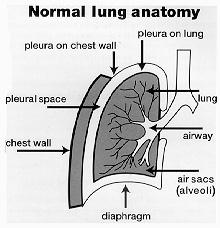
Mesothelioma is a rare form of cancer (malignancy) that most frequently arises from the cells lining the sacs of the chest (the pleura) or the abdomen (the peritoneum). Pleural mesothelioma is the most common form, often presenting with symptoms in the chest area. Peritoneal mesothelioma is much less common. This can effect the organs in the abdomen, and its symptoms are related to this area of the body, that is, abdominal swelling, nausea, vomiting, and bowel obstruction. The rarest form of mesothelioma is pericardial mesothelioma, which involves the sac surrounding the heart.
There are two major cell types of mesothelioma, epithelial and sarcomatoid. Sometimes both of these cell types can be present. The sarcomatoid type is rarer and occurs in only about 15% of cases; it portends a poorer prognosis. In very rare cases, mesothelioma can originate from benign, non-malignant cells. This so-called benign mesothelioma can be cured surgically.
As the disease progresses, shortness of breath increases, and weight loss, decreased appetite, and night sweats can develop. Local invasion by the tumor can result in changing of voice, loss of function of the diaphragm, and symptoms specific to the area and involvement of adjacent structures.
What causes mesothelioma?
Most people with malignant mesothelioma have worked on jobs where they breathed asbestos. Usually, this involves men over 40 years of age. Others have been exposed to asbestos in a household environment, often without knowing it. Interestingly, the number of new cases of mesothelioma has been relatively stable since 1983, the same time that the restrictions on asbestos were instituted by the U.S. Occupational Safety and Health Administration (OSHA). In Europe, the number of new cases of mesothelioma continues to rise.
Mesothelioma is a rare form of cancer (malignancy) that most frequently arises from the cells lining the sacs of the chest (the pleura) or the abdomen (the peritoneum). Pleural mesothelioma is the most common form, often presenting with symptoms in the chest area. Peritoneal mesothelioma is much less common. This can effect the organs in the abdomen, and its symptoms are related to this area of the body, that is, abdominal swelling, nausea, vomiting, and bowel obstruction. The rarest form of mesothelioma is pericardial mesothelioma, which involves the sac surrounding the heart.
There are two major cell types of mesothelioma, epithelial and sarcomatoid. Sometimes both of these cell types can be present. The sarcomatoid type is rarer and occurs in only about 15% of cases; it portends a poorer prognosis. In very rare cases, mesothelioma can originate from benign, non-malignant cells. This so-called benign mesothelioma can be cured surgically.
What are the symptoms of mesothelioma?
Most people present with complaints of shortness of breath. They also can have complaints of chest pain. Surprisingly, this pain is often not pleuritic; that is, it does not get worse with deep breathing. This is surprising in that the pleura (outer surface of the lung) is often involved in this disease, and most other diseases involving the pleura are often associated with pleuritic pain (pain that worsens with deep breathing). Patients may also be asymptomatic, with the disease discovered by physical exam or an abnormal chest X-ray.As the disease progresses, shortness of breath increases, and weight loss, decreased appetite, and night sweats can develop. Local invasion by the tumor can result in changing of voice, loss of function of the diaphragm, and symptoms specific to the area and involvement of adjacent structures.
What causes mesothelioma?
Most people with malignant mesothelioma have worked on jobs where they breathed asbestos. Usually, this involves men over 40 years of age. Others have been exposed to asbestos in a household environment, often without knowing it. Interestingly, the number of new cases of mesothelioma has been relatively stable since 1983, the same time that the restrictions on asbestos were instituted by the U.S. Occupational Safety and Health Administration (OSHA). In Europe, the number of new cases of mesothelioma continues to rise.
Wednesday, June 3, 2009
Balance for children
BCA Balance today : Rp. 400.000
No. Account BCA : 428 1320 429
Name : Steven Leonardo
No. Account BCA : 428 1320 429
Name : Steven Leonardo
Subscribe to:
Posts (Atom)